Chromosomes
DNA is not stored in the cell as a tangled heap, but packed into “volumes” called chromosomes. In leukemia, breaks and rearrangements in these volumes are often the key to the diagnosis, the risk group and even the treatment.

01Short answer
02Simple explanation
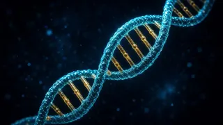
Imagine DNA as an extremely long thread — unwound, the DNA from a single cell would be about two metres long. To stop it tangling, the cell winds it onto “spools” and folds it into compact packages — chromosomes. You can also think of chromosomes as the volumes of an encyclopedia.
Humans normally have 46 chromosomes, or 23 pairs. In each pair, one chromosome comes from the mother and one from the father. Twenty-two pairs are the same in everyone and are numbered (1 to 22, roughly from largest to smallest), while the 23rd pair determines sex: XX in women, XY in men.
In a leukemia cell, these volumes can have “binding errors”. A piece of one volume can break off and attach to another — this is a translocation. Sometimes a piece is lost (a deletion), and sometimes a whole volume is present in an extra copy or goes missing entirely.
The best-known example is the Philadelphia chromosome: chromosomes 9 and 22 swap pieces, and at the break point two genes fuse into a new “hybrid” gene — BCR::ABL1. It makes a protein that drives the cell to divide non-stop. The Philadelphia chromosome is found in almost all cases of CML and in some adults with ALL. Drugs that block it have transformed treatment.
These changes were not passed on at birth — they arose only in the leukemia cells. To find them, doctors examine the chromosomes in a bone marrow or blood sample. The result helps pin down the exact name of the leukemia, the risk group and the choice of treatment.
03Scientific explanation
Each chromosome consists of DNA wound around histone proteins (chromatin). A constriction called the centromere divides it into a short (p) arm and a long (q) arm, with protective telomeres at the ends. Before a cell divides, each chromosome is copied; at metaphase the chromosomes are at their most compact and can be seen under the microscope. In cytogenetic notation, “t(9;22)(q34;q11)” means a translocation between region q34 of chromosome 9 and q11 of chromosome 22; “del(5q)” means loss of part of the long arm of chromosome 5; “+8” an extra chromosome 8; and “−7” loss of chromosome 7.
Chromosome changes act in leukemia in two main ways. Balanced translocations and inversions join two genes into a fusion gene (BCR::ABL1, PML::RARA, RUNX1::RUNX1T1, CBFB::MYH11, ETV6::RUNX1, KMT2A fusions) or move a gene next to a strong regulator that boosts its activity. Numerical changes and deletions (e.g. hyperdiploidy, hypodiploidy, −7, del(5q), del(17p)) change the “dose” of genes or remove guardian genes such as TP53.
These changes are at the heart of classification. In AML, t(15;17), t(8;21) and inv(16)/t(16;16) are defining abnormalities, and when present the diagnosis can be made with fewer than 20% blasts; t(8;21) and inv(16) are generally in the favourable group, while a complex karyotype (several abnormalities), −7 and some other changes are in the less favourable group (ELN 2022). In childhood B-ALL, high hyperdiploidy (51–65 chromosomes) and ETV6::RUNX1 are generally favourable, while BCR::ABL1, KMT2A::AFF1 and hypodiploidy (<44 chromosomes) are less favourable. In CLL, FISH looks for del(13q), +12, del(11q) and del(17p): isolated del(13q) is favourable, while del(17p) is a high-risk feature that affects treatment choice.
The main tests are karyotyping (a G-banded survey of all chromosomes; it needs dividing cells, and usually at least 20 metaphases are analysed), FISH (fluorescent probes that look for specific changes; it works on non-dividing cells and gives faster results) and molecular methods (RT-PCR, NGS and, in some centres, optical genome mapping). When APL is suspected, urgent confirmation of PML::RARA is especially important.
04What happens in the body?
Translocation: two chromosomes swap pieces
When a cell divides, two chromosomes break and their pieces change places. At the break point, two genes can fuse into a new fusion gene. This is how the Philadelphia chromosome (BCR::ABL1) and PML::RARA in APL arise. These fusions are both a diagnostic “fingerprint” and a treatment target.

Extra or missing chromosomes
Some leukemia cells have a changed number of chromosomes. In childhood ALL, having many extra chromosomes (high hyperdiploidy) is usually a good sign. Having fewer chromosomes (hypodiploidy) or losing chromosome 7 may call for more intensive treatment.
How do karyotyping and FISH differ?
A karyotype is a “group photo” of all the chromosomes: it shows changes in number and large structural rearrangements, but it needs dividing cells and can take days to weeks. FISH looks for a specific change using fluorescent “lights”; it is faster, but finds only what it is looking for.

What does my result tell me?
Chromosome findings point to the leukemia subtype, the risk group and sometimes whether a targeted drug is suitable. No single finding decides everything — your doctor weighs it alongside other tests, response to treatment and MRD.
05From chromosome to treatment decision
A chromosome change shapes your doctor’s decision in four steps.



06Common chromosome changes in leukemia
| Change | Where is it found? | What happens? | What does it usually mean? |
|---|---|---|---|
| t(9;22) — Philadelphia chromosome | CML; some ALL (more often in adults) | BCR::ABL1 fusion gene | Targeted drugs (TKIs) work |
| t(15;17) | APL | PML::RARA fusion gene | Very good response to specific treatment |
| t(8;21), inv(16) | AML | RUNX1::RUNX1T1, CBFB::MYH11 | Usually favourable |
| High hyperdiploidy (51–65 chromosomes) | Childhood B-ALL | Extra chromosomes | Usually favourable |
| t(12;21) | Childhood B-ALL | ETV6::RUNX1 fusion gene | Usually favourable |
| KMT2A (11q23) rearrangements | Infant ALL, AML | KMT2A fusions | Often less favourable |
| Hypodiploidy (<44 chromosomes) | ALL | Loss of chromosomes | Less favourable |
| Complex karyotype, −7, del(5q) | AML, MDS | Multiple losses and rearrangements | Less favourable |
| Isolated del(13q) | CLL | Loss of part of chromosome 13 | Usually favourable |
| del(17p) | CLL and others | Loss of the TP53 gene | High risk; affects treatment choice |
* Outlook depends not on a single finding but on all features together, age, response to treatment and MRD. New drugs are changing the meaning of some changes. Discuss your results with your doctor.
07Key terms
- Chromosome
- A “volume” of DNA tightly wound around proteins.
- Karyotype
- The number and appearance of all of a cell’s chromosomes; also the test that shows them.
- Autosome
- Chromosomes 1–22, which are not related to sex.
- Centromere
- The constriction dividing a chromosome into short (p) and long (q) arms.
- Translocation
- An exchange of pieces between two chromosomes.
- Deletion
- Loss of part of a chromosome.
- Inversion
- A piece of chromosome breaks off and reattaches upside down.
- Fusion gene
- A new gene formed by joining two different genes.
- FISH
- A test that finds specific chromosome changes using fluorescent probes.
- Philadelphia chromosome
- The shortened chromosome 22 resulting from t(9;22); it carries BCR::ABL1.
08Key points
- Chromosomes are compact packages of DNA; humans normally have 46 (23 pairs).
- In leukemia cells, chromosomes can break, swap pieces, be lost or be duplicated.
- A translocation can join two genes into a fusion gene (e.g. BCR::ABL1).
- These changes are usually only in the leukemia cells and are not inherited.
- A karyotype shows the overall picture; FISH shows specific changes.
- Chromosome findings shape the diagnosis, the risk group and the choice of treatment.
- Targeted drugs against the Philadelphia chromosome are one of the great successes of modern leukemia treatment.
09Frequently asked questions
Are the chromosome changes in leukemia cells inherited?
Usually not. These changes arise during life, only in blood-forming cells, and are not present in the body’s other cells, so they are not passed on to children. The exception is chromosome conditions present from birth, such as Down syndrome, which can raise the risk of leukemia.
Why do the results take so long?
For a karyotype, the cells have to be grown in the lab for a while and then the chromosomes analysed one by one — this takes several days, sometimes longer. FISH and some molecular tests are quicker. In urgent situations (such as suspected APL), doctors prioritise the most important tests.
My karyotype is “normal”. Is that good or bad?
In AML, a normal karyotype is common and usually falls into the intermediate risk group; the final picture depends on molecular tests (such as NPM1, FLT3 and CEBPA). So a “normal karyotype” on its own is neither good nor bad news — it is one part of the picture.
Are chromosome tests repeated after treatment?
Sometimes, yes. Checking whether a change found at diagnosis has disappeared helps assess the response to treatment. In CML, for example, BCR::ABL1 levels are tracked regularly with a molecular test.
My report says “46,XY” or “46,XX”. What does that mean?
It means the cells examined had 46 chromosomes and shows the sex chromosomes (XY — male, XX — female). If nothing else follows, no visible chromosome change was found in the cells examined. Either way, go through the result with your doctor.
Is the Philadelphia chromosome bad news?
Today the Philadelphia chromosome is also an “opportunity”: there are drugs that block it. Thanks to them, most people with CML live long, active lives. In Philadelphia-positive ALL, these drugs are added to treatment and have greatly improved results.
10Questions for your doctor
Take these questions to your appointment. The ones you save are collected on the “My questions” page and stay on this device only.
Which chromosome changes did the karyotype or FISH show?
Which risk group does this change put me in?
Is this change inherited, and does it affect my family?
Will you check for this change again during treatment?
12Sources
- MedlinePlus Genetics (U.S. National Library of Medicine). What is a chromosome?
- MedlinePlus Genetics (U.S. National Library of Medicine). Can changes in the number or structure of chromosomes affect health and development?
- National Human Genome Research Institute (NHGRI). Chromosomes Fact Sheet
- National Human Genome Research Institute (NHGRI). Translocation — Talking Glossary
- National Cancer Institute (NCI). NCI Dictionary of Cancer Terms: Philadelphia chromosome
- National Cancer Institute (NCI). Adult Acute Myeloid Leukemia Treatment (PDQ®) — Health Professional Version
- National Cancer Institute (NCI). Childhood Acute Lymphoblastic Leukemia Treatment (PDQ®) — Health Professional Version
- National Cancer Institute (NCI). Chronic Lymphocytic Leukemia Treatment (PDQ®) — Health Professional Version
- National Cancer Institute (NCI). Childhood Acute Lymphoblastic Leukemia Treatment (PDQ®) — Patient Version
- American Cancer Society. How Is Acute Myeloid Leukemia Classified?
- American Cancer Society. Typing of Acute Lymphocytic Leukemia (ALL)
- Leukemia (Nature) — WHO 5th edition. The 5th edition of the World Health Organization Classification of Haematolymphoid Tumours: Myeloid and Histiocytic/Dendritic Neoplasms