Other genetic changes
The Philadelphia chromosome is the best-known genetic change in leukemia, but far from the only one. Reports may mention NPM1, FLT3, TP53, KMT2A, ETV6::RUNX1 or del(17p). This topic calmly explains the most common of these other changes, the kinds they come in and what they mean for treatment.

01Short answer
02Simple explanation
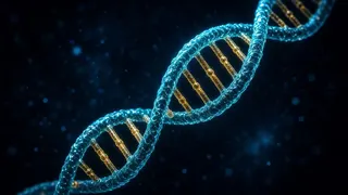
Think of genes as the cell’s instruction manual. In leukemia some pages of this manual have changed: some have swapped places, some are missing or duplicated, and some contain a typo.
A fusion gene forms when pieces of two chromosomes swap places: parts of two different genes join, creating a new gene that works wrongly.
A point mutation is a small “typo” in a gene. Names like NPM1, FLT3 and TP53 refer to changes of this kind.
Some changes are linked to a more favourable course, some to a more difficult one, and some show that a particular targeted drug is likely to work.
Most of these changes are not inherited: they arise only in the leukemia cells and are not passed on to your children.
03Scientific explanation
AML. The WHO 2022 and ICC classifications name many types of AML after their genetic change: RUNX1::RUNX1T1 [t(8;21)], CBFB::MYH11 [inv(16)], PML::RARA [t(15;17)], KMT2A rearrangements, NPM1 mutation, CEBPA mutation and others. In the ELN 2022 risk system, for example, t(8;21), inv(16) and NPM1 mutation without FLT3-ITD are favourable, while TP53 mutation, a complex karyotype, monosomy 7, some KMT2A rearrangements and mutations in genes such as ASXL1 and RUNX1 are adverse.
ALL. According to the ACS, in childhood B-ALL, ETV6::RUNX1 and high hyperdiploidy (many extra chromosomes) are more favourable, while KMT2A rearrangements, hypodiploidy, iAMP21 and TCF3::HLF are linked to a more difficult course. In “Ph-like” ALL there is no Philadelphia chromosome, but there are similar signalling changes, and targeted drugs are being studied for some of them.
CLL. According to the NCI, FISH is used to check for del(13q), trisomy 12, del(11q) and del(17p), and molecular testing for TP53 mutation and IGHV mutation status. With del(17p) or a TP53 mutation, chemoimmunotherapy usually works less well and targeted drugs are preferred.
Somatic and inherited (germline). The NCI explains that somatic changes arise during life in individual cells and are not inherited, whereas germline changes are present in every cell of the body and can run in families. Inherited predisposition is rare in leukemia, but it matters when several relatives have had blood disorders, or when choosing a related donor.
04What happens in the body?
Three kinds of change
Translocations and fusion genes (pieces of two chromosomes swap places), changes in chromosome number (an extra or missing chromosome) and point mutations (a small change in one gene). Each is found with its own test: karyotype, FISH, PCR or NGS.
Fusion genes
When two chromosomes break and swap pieces, parts of two different genes end up side by side and a new “hybrid” gene forms. It gives the cell wrong instructions — for example, stopping maturation or speeding up division. Fusion genes often appear in the name of the diagnosis itself.

Point mutations
A change in one or a few “letters” of a gene, or a small insertion or loss. An NGS panel checks dozens of genes at once. The report gives the gene name, the type of change and the variant allele frequency (VAF) — the percentage of DNA in which the mutation was found.

How does this change treatment?
Genetics defines the risk group: in AML, for example, an adverse risk may lead to considering a transplant earlier. Some mutations are targets for specific drugs: FLT3 inhibitors, IDH1 and IDH2 inhibitors and, in some situations with KMT2A or NPM1 changes, menin inhibitors.
What does it mean for the family?
Usually nothing: the change is only in the leukemia cells. But if several relatives have had blood disorders, the doctor may offer a test on non-leukemia tissue (such as skin or hair follicles) and genetic counselling. This also matters when choosing a donor among relatives.
05From a genetic finding to a decision
A small change, a lot of information.



06Some genetic changes and their general meaning
| Change | Where it occurs | General meaning |
|---|---|---|
| RUNX1::RUNX1T1, CBFB::MYH11 | AML | Usually favourable risk (ELN 2022) |
| NPM1 (without FLT3-ITD) | AML | Usually favourable risk |
| FLT3 (ITD, TKD) | AML | Target for FLT3 inhibitors |
| IDH1, IDH2 | AML | Target for IDH inhibitors |
| TP53, complex karyotype | AML, ALL, CLL | More difficult course; a specific approach |
| ETV6::RUNX1, high hyperdiploidy | Childhood B-ALL | Usually favourable |
| KMT2A rearrangements | AML, ALL (especially in infants) | Often high risk; new targeted options being studied |
| del(17p), unmutated IGHV | CLL | Change the choice of treatment |
* This is a simplified general table. What a change means for you depends on all the results taken together and can only be explained by your hematologist.
07Key terms
- Translocation
- Two chromosomes exchanging pieces.
- Fusion gene
- A new gene formed by joining parts of two genes.
- Point mutation
- A change in one or a few “letters” of a gene.
- Complex karyotype
- Three or more chromosome changes at the same time.
- Hyperdiploidy
- More chromosomes in a cell than normal.
- NGS
- Next-generation sequencing, which reads many genes at once.
- VAF
- Variant allele frequency — the percentage of DNA carrying the mutation.
- Somatic change
- A change acquired during life that is not inherited.
- Germline change
- A change present in all body cells that can run in families.
08Key points
- The Philadelphia chromosome is not the only important genetic change.
- Changes come as translocations, changes in chromosome number and point mutations.
- Genetics defines the subtype, the risk group and targeted drugs.
- A single gene does not decide the outlook on its own.
- Most changes are not inherited.
- If several relatives have had blood disorders, tell your doctor.
- Always discuss genetic results with your hematologist.
09Frequently asked questions
My report says “TP53 mutation”. What does that mean?
TP53 is an important gene that protects cells from damage. A mutation in it is linked to a more difficult course of leukemia and a weaker response to some drugs. But it does not mean there are no treatment options — your doctor will explain the approach that suits you.
Why do genetic results take so long?
For a karyotype, cells have to be grown in the laboratory; for NGS, complex reading and analysis are needed. Tests that are needed urgently (for example FLT3 and PML::RARA) are often done faster.
Do my children carry this change?
Usually not: most changes in leukemia are present only in the leukemia cells. If an inherited predisposition is suspected, your doctor will offer a specific test and genetic counselling.
Can the genetics change during treatment?
Yes, leukemia cells can acquire new changes over time, especially at relapse. That is why tests are often repeated at relapse.
What does “VAF 40%” mean?
It means the mutation was found in roughly 40% of the DNA in the sample. The number depends on the proportion of leukemia cells and other factors; your doctor can explain what it means.
10Questions for your doctor
Take these questions to your appointment. The ones you save are collected on the “My questions” page and stay on this device only.
My report says “TP53 mutation”. What does that mean?
Why do genetic results take so long?
Do my children carry this change?
Can the genetics change during treatment?
What does “VAF 40%” mean?
12Sources
- American Cancer Society. How Is Acute Myeloid Leukemia Classified?
- American Cancer Society. Typing of Acute Lymphocytic Leukemia (ALL)
- American Cancer Society. Genes and Cancer
- National Cancer Institute (NCI). The Genetics of Cancer
- National Cancer Institute (NCI). Biomarker Testing for Cancer Treatment
- National Cancer Institute (NCI). NCI Dictionary of Cancer Terms: germline mutation
- National Cancer Institute (NCI). NCI Dictionary of Cancer Terms: translocation
- MedlinePlus Genetics (U.S. National Library of Medicine). What is a gene variant and how do variants occur?
- MedlinePlus Genetics (U.S. National Library of Medicine). Can changes in the number or structure of chromosomes affect health and development?
- Blood (American Society of Hematology), 2022. Dohner H, et al. Diagnosis and management of AML in adults: 2022 recommendations from an international expert panel on behalf of the ELN
- Leukemia (journal), 2022. Khoury JD et al. The 5th edition of the WHO Classification of Haematolymphoid Tumours: Myeloid and Histiocytic/Dendritic Neoplasms
- Leukemia (journal), 2022. Alaggio R et al. The 5th edition of the WHO Classification of Haematolymphoid Tumours: Lymphoid Neoplasms
- National Cancer Institute (NCI). Chronic Lymphocytic Leukemia Treatment (PDQ®) — Health Professional Version