How leukemia subtypes are identified
“AML”, “B-ALL”, “M2”, “t(9;22)”: all these words in discharge summaries describe the leukemia subtype. This topic explains which tests doctors use and how they reason to identify the subtype.

01Short answer
02Simple explanation
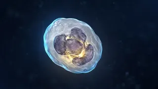
Appearance. A specialist looks at cell size, nucleus shape and the granules inside. This gives the first clue.
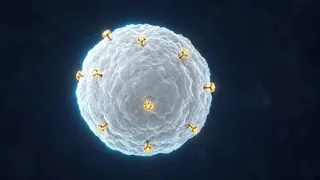
“Markers”. Every cell carries protein markers on its surface called CDs. For example, CD19 is typical of B cells, CD3 of T cells, and CD33 and MPO of myeloid cells.
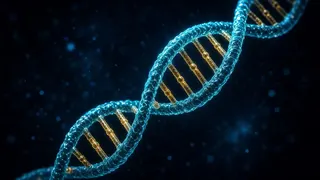
Genetics. Some chromosome changes are so characteristic that the subtype is named after them, for example “AML with inv(16)”.
Putting it together. All results are assessed together and given a final name according to classification rules.
03Scientific explanation
In AML: according to the ACS, the FAB system divided AML into M0–M7 by cell appearance (for example, M3 is APL and M4 is myelomonocytic). The WHO and ICC systems rely first on genetics: NPM1, CEBPA, RUNX1::RUNX1T1, CBFB::MYH11, PML::RARA and others.
In ALL: according to the ACS, ALL is first divided by immunophenotype into B-ALL and T-ALL, and B-ALL is then split into genetic subgroups: BCR::ABL1 (Philadelphia chromosome), KMT2A rearrangements, hyperdiploidy, ETV6::RUNX1 and others.
Mixed phenotype: rarely, cells carry both myeloid and lymphoid markers (MPAL). This needs a special approach.
04What happens in the body?
Why so many tests?
Two leukemias that look alike under the microscope can have completely different genetics and treatment. Only the combination of all tests gives the right subtype.

What might your discharge summary say?
For example: “B-ALL, immunophenotype: CD19+, CD10+, CD22+, TdT+, cytoplasmic CD79a+; karyotype: hyperdiploid”. This means ALL from the B-cell family, made of immature (TdT+) cells, with an increased number of chromosomes.

Can the subtype change?
The core subtype usually stays the same, but new genetic changes can appear at relapse. That is why tests are repeated if the leukemia returns.
05Four views of a subtype
Each test adds part of the picture.


06Tests that define the subtype
| Test | What does it look at? | Example result |
|---|---|---|
| Morphology | Cell shape, granules | Blasts with maturation (M2) |
| Cytochemistry | Enzyme stains | MPO positive |
| Flow cytometry | CD markers | CD19+, CD10+ (B-ALL) |
| Karyotype / FISH | Chromosomes | t(15;17): APL |
| Molecular tests | Gene mutations | NPM1, FLT3-ITD |
* Examples are for illustration; your result may differ.
07Key terms
- Morphology
- Studying how cells look under a microscope.
- CD marker
- A recognition protein on the cell surface.
- FAB
- An older classification based on appearance.
- WHO / ICC
- Modern classifications that include genetics.
- MPAL
- Leukemia with both myeloid and lymphoid markers.
08Key points
- A subtype comes from three sources of data.
- Morphology is the first clue.
- CD markers show the cell family.
- Genetics often gives the final name.
- FAB is older; WHO/ICC are current.
- The subtype guides treatment.
09Frequently asked questions
My summary says “M2”. What does that mean?
It is AML with maturation in the FAB system. Today it is assessed together with the genetic result.
Does the subtype decide the outlook?
It is one factor; others are genetics, response to treatment and general health.
Why are there two classifications (WHO and ICC)?
In 2022 two expert groups produced classifications in parallel. They are very similar and differ mostly in details.
Can a subtype be wrongly identified?
Rarely; that is why results are checked with several methods and a second opinion is sought when needed.
10Questions for your doctor
Take these questions to your appointment. The ones you save are collected on the “My questions” page and stay on this device only.
My summary says “M2”. What does that mean?
Does the subtype decide the outlook?
Why are there two classifications (WHO and ICC)?
Can a subtype be wrongly identified?
12Sources
- American Cancer Society. How Is Acute Myeloid Leukemia Classified?
- American Cancer Society. How Is Acute Lymphocytic Leukemia Classified?
- National Cancer Institute (NCI). Adult Acute Myeloid Leukemia Treatment (PDQ) – Patient Version
- MedlinePlus. Flow cytometry
- Cancer Research UK. Types of acute myeloid leukaemia (AML)
- World Health Organization / IARC. WHO Classification of Tumours: Haematolymphoid Tumours, 5th edition (2022)
- ECOG-ACRIN Cancer Research Group. ECOG Performance Status Scale
- American Cancer Society. Key Statistics for Childhood Leukemia
- National Institute for Health and Care Excellence (NICE). Suspected cancer: recognition and referral (NG12)
- Children & Young People’s Cancer Association (CCLG). Leukaemia in children