Biomarkers: CD markers, genetic markers and MRD
Test reports after a leukemia diagnosis are often full of puzzling letters and numbers: CD19+, CD33, FLT3-ITD, NPM1, BCR::ABL1, “MRD 0.01%”. These are biomarkers — a kind of “ID card” for the leukemia cell. This topic explains the main types of biomarkers, how they are measured and what they mean for diagnosis, choosing treatment and tracking the response to it.

01Short answer
02Simple explanation
A biomarker is a “clue” that tells us something about a disease — a protein on the surface of a cell, a change in DNA or a substance in the blood. In leukemia, biomarkers help the doctor understand the exact type of disease, how aggressive it may be and which medicine is likely to work best.
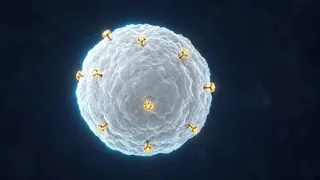
CD markers are “flags” on the surface of a cell. Each type of cell has its own set of flags. The laboratory finds them with special glowing antibodies and works out which cell the leukemia came from — a myeloid cell, a B lymphocyte or a T lymphocyte. A “+” means the marker is present; a “−” means it is absent.
Genetic markers are changes in the DNA of the leukemia cell: pieces of chromosomes swapping places, two genes fusing together or a small error in a single gene. Some changes improve the outlook, others call for more intensive treatment, and some have dedicated targeted drugs.
MRD (measurable residual disease) is the very small number of leukemia cells left in the body after remission is reached. They cannot be seen under the microscope, but sensitive tests can find them. MRD shows how deeply treatment has worked and helps plan the next steps.
Biomarker results are not read one by one but together. A single marker rarely tells the whole story. Do not hesitate to ask your hematologist what your results mean — it is a completely normal and important question.
03Scientific explanation
Immunophenotyping is a method that identifies cells using antibodies that bind to antigens (markers) on their surface (NCI); it is usually done with a flow cytometer. Examples: CD34 is found on immature (blast) cells; B-ALL typically shows CD19, CD10, CD22 and TdT, and T-ALL shows CD2, CD3 and CD7; AML carries myeloid markers such as CD33, CD13 and CD117, as well as myeloperoxidase. CLL is typically made of B cells carrying CD5 and CD23, while hairy cell leukemia typically shows CD11c, CD25, CD103 and CD123 (NCI). Some markers are also treatment targets: CD33 for gemtuzumab ozogamicin, CD19 for blinatumomab and CAR-T cells, CD22 for inotuzumab ozogamicin, and CD20 for anti-CD20 antibodies in CLL (rituximab, obinutuzumab).
Genetic markers are found with karyotyping, FISH, PCR and next-generation sequencing (NGS). They refine the diagnosis and define the risk group: for example, ELN 2022 places an NPM1 mutation (without FLT3-ITD) in AML in the favourable group and a TP53 mutation in the adverse group. Examples of targeted therapy: BCR::ABL1 — tyrosine kinase inhibitors (TKIs); PML::RARA — ATRA and arsenic trioxide; FLT3 — midostaurin, gilteritinib, quizartinib; IDH1 — ivosidenib; IDH2 — enasidenib; NPM1 mutations and KMT2A rearrangements — menin inhibitors. The NCI points out that biomarker testing of a cancer is different from genetic testing for inherited mutations.
Measuring MRD: according to the NCI, in childhood ALL flow cytometry or PCR can detect 1 leukemia cell among 100,000 normal cells, and a level of 1 in 10,000 (0.01%) is detected routinely; high-throughput sequencing of immunoglobulin and T-cell receptor gene rearrangements raises the sensitivity to 1 in 1,000,000 (0.0001%). MRD at the end of induction is an important independent predictor of outcome in children and adolescents with B-ALL. In AML, the 2021 ELN consensus sets flow-cytometry MRD positivity at 0.1% or more, and recommends quantitative PCR for NPM1-mutated AML.
MRD in treatment decisions: the FDA has approved blinatumomab for adults and children with B-ALL in first or second complete remission with MRD of 0.1% or more. In CML, response is tracked by measuring BCR::ABL1 with PCR on the International Scale (IS): ≤10% at 3 months is an early response, ≤0.1% a major molecular response (MMR), ≤0.01% MR4 and ≤0.0032% MR4.5. In CLL, undetectable MRD after fixed-duration treatment is being studied as a measure of how deep the response is. A negative MRD result is not a guarantee of cure, and a positive one does not mean relapse is inevitable.
04What happens in the body?
The cell surface: CD markers
In flow cytometry, cells pass one at a time in a thin stream in front of a laser beam. The antibodies attached to them glow in different colours, and the instrument records which markers each cell carries — thousands of cells per second. This way, within hours or days, it becomes clear which cell line the leukemia belongs to.

Inside the cell: genetic markers
Chromosome analysis, FISH and molecular tests show what has changed in the DNA of the leukemia cell. Some results come back quickly, while others take longer. Sometimes doctors wait for certain genetic results before starting treatment because they can change the choice of drugs; in urgent situations, treatment starts without waiting for them.
Measuring the invisible: MRD
In remission, no leukemia cells can be seen under the microscope, yet a few may still remain. An MRD test counts these leftovers using flow cytometry, PCR or sequencing. MRD is checked at set points during treatment, and its trend — falling or rising — says more than any single measurement.

Results are read together
Your hematologist weighs CD markers, genetic findings, blood tests, age and general health together. The same marker can mean different things in different types of leukemia. That is why it is better to discuss results with your doctor than to look them up one by one online.
05Biomarkers along the treatment path
Four steps — from the sample to tracking the response to treatment.




06Common biomarkers: a quick guide
| Marker | Typical in | What it shows | Link to treatment |
|---|---|---|---|
| CD34 | AML and ALL blasts | An immature cell | Helps recognise blasts |
| CD33, CD13, CD117 | AML | Myeloid line | CD33 is the target of gemtuzumab ozogamicin |
| CD19, CD10, CD22, TdT | B-ALL | B-cell precursor | CD19: blinatumomab, CAR-T; CD22: inotuzumab |
| CD2, CD3, CD7 | T-ALL | T-cell line | Diagnosis and MRD tracking |
| CD5, CD23, CD20 | CLL | Typical CLL pattern | CD20 is the target of anti-CD20 antibodies |
| CD11c, CD25, CD103, CD123 | Hairy cell leukemia | Typical HCL pattern | Confirms the diagnosis |
| BCR::ABL1 | CML, some ALL | Fusion gene | TKIs; monitoring by PCR |
| PML::RARA | APL | Fusion gene | ATRA + arsenic trioxide |
| FLT3, IDH1/IDH2, NPM1 | AML | Gene mutations | FLT3, IDH and menin inhibitors |
| TP53 / del(17p), IGHV | CLL | Outlook | Choice of treatment type |
* This table is a simplified summary: markers can appear in different combinations in different leukemias, and the choice of treatment depends on many factors. Discuss your results with your hematologist.
07Key terms
- Biomarker
- A measurable biological substance that is a sign of a normal or abnormal process.
- CD marker
- A numbered protein on the cell surface (“cluster of differentiation”).
- Immunophenotyping
- Identifying cells by their surface markers using antibodies.
- Flow cytometry
- A method that measures cell markers by passing cells in front of a laser beam.
- Fusion gene
- An abnormal gene formed when parts of two different genes join, such as BCR::ABL1.
- FISH
- A test that finds specific chromosome changes using fluorescent probes.
- NGS
- Next-generation sequencing — a test that reads many genes at once.
- MRD
- Measurable residual disease — a small number of leukemia cells left after treatment.
- International Scale (IS)
- A standard for comparing BCR::ABL1 levels in CML between laboratories.
- Targeted therapy
- A drug aimed at a specific molecule in the leukemia cell.
08Key points
- Biomarkers are the leukemia’s “ID card”.
- CD markers are measured by flow cytometry and show the leukemia’s cell line.
- Some CD markers are targets for antibody drugs and CAR-T therapy.
- Genetic markers define the type, risk group and targeted drugs.
- Biomarker testing of a cancer is different from inherited genetic testing.
- MRD measures invisible residual disease — down to 1 cell in a million.
- A negative MRD is not a guarantee, and a positive one is not a verdict.
- Results should always be read together and with your doctor.
09Frequently asked questions
My report says “CD19+”. What does that mean?
It means the leukemia cells carry the CD19 marker on their surface, which is typical of B cells. It helps confirm the diagnosis and can be a target for some immunotherapies. Only your hematologist, looking at all the results, can say which treatment is right for you.
Will my genetic markers be passed on to my children?
Usually not. The changes found in leukemia cells (such as FLT3 or BCR::ABL1) are acquired during life and are not inherited. If an inherited predisposition is suspected, a separate genetic test and counselling are offered.
Why do doctors sometimes wait for genetic results before starting treatment?
Some results can change the choice of drugs — for example, in APL or with a FLT3 mutation. But if your condition needs urgent treatment, doctors may start without waiting and refine the plan afterwards.
If my MRD is negative, am I cured?
A negative MRD is a very good sign, but it cannot guarantee that no cells remain below the test’s detection limit. That is why the treatment plan is usually completed in full and follow-up continues.
If my MRD is positive, will I definitely relapse?
No. A positive MRD adds information about risk and often becomes the reason to intensify or change treatment. For example, there are treatments specifically approved for MRD-positive B-ALL.
How often are these tests repeated?
That depends on the type of leukemia and the treatment protocol. For example, in CML the BCR::ABL1 level is measured regularly in the blood, while in acute leukemias MRD is checked at particular stages of treatment. Your hematologist will explain the schedule.
10Questions for your doctor
Take these questions to your appointment. The ones you save are collected on the “My questions” page and stay on this device only.
Which CD markers and genetic markers were found in my case?
Which method will you use to measure MRD, and when?
How could my MRD result change my treatment plan?
Can I have a copy of my biomarker results?
12Sources
- National Cancer Institute (NCI). Biomarker Testing for Cancer Treatment
- National Cancer Institute (NCI). NCI Dictionary of Cancer Terms: minimal residual disease
- National Cancer Institute (NCI). NCI Dictionary of Cancer Terms: immunophenotyping
- National Cancer Institute (NCI). Childhood Acute Lymphoblastic Leukemia Treatment (PDQ®) — Health Professional Version
- National Cancer Institute (NCI). Adult Acute Myeloid Leukemia Treatment (PDQ®) — Health Professional Version
- National Cancer Institute (NCI). Chronic Lymphocytic Leukemia Treatment (PDQ®) — Health Professional Version
- National Cancer Institute (NCI). Chronic Myeloid Leukemia Treatment (PDQ®) — Health Professional Version
- National Cancer Institute (NCI). Hairy Cell Leukemia Treatment (PDQ®) — Health Professional Version
- National Cancer Institute (NCI). Targeted Therapy to Treat Cancer
- Blood (American Society of Hematology), 2021. 2021 Update on MRD in acute myeloid leukemia: a consensus document from the European LeukemiaNet MRD Working Party
- Blood (American Society of Hematology), 2022. Diagnosis and management of AML in adults: 2022 recommendations from an international expert panel on behalf of the ELN
- U.S. Food and Drug Administration (FDA). FDA expands approval of Blincyto for treatment of a type of leukemia in patients who have a certain risk factor for relapse
- American Cancer Society. How Is Acute Myeloid Leukemia Classified?
- American Cancer Society. Typing of Acute Lymphocytic Leukemia (ALL)