Cell division and cell death
In a healthy body there is a fine balance between making new cells and removing old ones. Leukemia is a breakdown of that balance: cells divide more than they should and do not die when they should. This topic helps explain the logic of both the disease and its treatment.

01Short answer
02Simple explanation
Your body is constantly renewing itself. The bone marrow makes billions of new blood cells every day — and roughly as many old cells die. For numbers to stay steady, birth and death have to balance each other.
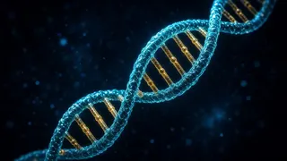
Before dividing, a cell copies its entire DNA “book”, then splits in two, and each new cell receives a complete copy. This is called mitosis. The process is not random: a cell divides only when it receives a “divide” signal and everything is in order.
Along the way there are checkpoints — like sentries. They ask: “Has the cell grown enough? Was the DNA copied correctly?” If an error is found, the cycle pauses and repair begins. If the error cannot be fixed, the cell sacrifices itself.
This selfless “self-shutdown” is called apoptosis. The cell neatly takes itself apart, and clean-up cells swallow the pieces — without inflammation or harm. Apoptosis protects the body from damaged and dangerous cells.
In leukemia, this system breaks down: the “divide” button sticks, the sentries fall silent and the “self-destruct button” stops working. As a result, abnormal cells build up. Treatment exploits these weak points: chemotherapy hits dividing cells, and some newer drugs restart apoptosis.
03Scientific explanation
The cell cycle has four phases: G1 (growth and preparation for division), S (DNA synthesis — the whole genome is copied), G2 (checking the copy and preparing for mitosis) and M (mitosis — separation of the chromosomes and cytokinesis). Non-dividing cells rest in G0; most blood-forming stem cells also spend much of their time in G0. Progress through the cycle is driven by cyclins and cyclin-dependent kinases (CDKs).
Checkpoints (G1/S, G2/M and the mitotic spindle checkpoint) catch errors. The best-known guardian is the p53 protein (TP53 gene): when DNA is damaged, it halts the cycle, starts repair and, if the damage is severe, triggers apoptosis. That is why losing TP53 gives a leukemia cell an advantage both in survival and in resisting chemotherapy. Telomeres at the ends of chromosomes shorten with each division and limit how many times a cell can divide; many cancer cells get around this limit.
Apoptosis is triggered by two main routes: the internal (mitochondrial) pathway, controlled by the BCL-2 family of proteins, and the external pathway, via “death receptors” on the cell surface. Both activate caspase enzymes, and the cell is dismantled in an orderly way. BCL-2 is a protein that blocks apoptosis; its activity is increased in CLL and in many cases of AML. Venetoclax blocks BCL-2 and restarts apoptosis; according to NCI, it is approved to treat CLL and some cases of AML.
Many chemotherapy drugs act at particular stages of the cycle: antimetabolites (cytarabine, methotrexate) disrupt DNA synthesis mainly in S phase, mitotic inhibitors (e.g. vincristine) stop chromosome separation in M phase, and anthracyclines and alkylating agents damage DNA and trigger apoptosis. Because these drugs also affect rapidly dividing normal cells — in the marrow, hair follicles and the lining of the mouth and gut — typical side effects occur. Leukemia cells “sleeping” in G0 may be less sensitive to chemotherapy; this is one possible reason for relapse and partly explains why treatment has several phases.
04What happens in the body?
The cell cycle: four stages
First the cell grows (G1), then it copies its DNA (S), checks the copy (G2) and finally divides in two (M). There is a checkpoint at the end of each stage. When there is no need to divide, the cell moves into a resting state (G0).

Apoptosis: a selfless death
Apoptosis is a cell’s orderly self-shutdown. It removes old and damaged cells without causing inflammation. Leukemia cells, however, often escape this process with the help of “protective” proteins such as BCL-2, and live longer than they should.

What goes wrong in leukemia?
Three things at once: the signal to divide stays on, maturation stops, and apoptosis fails. In acute leukemia, fast division and a maturation block dominate; in CLL, cells build up mainly because they live longer than they should.
How does treatment use this knowledge?
Chemotherapy targets rapidly dividing cells, which is why treatment is given in cycles with breaks, so that normal cells can recover. Targeted drugs switch off division signals, and drugs such as venetoclax restore apoptosis.
05The life of a cell
A healthy blood cell lives by a four-step programme. A leukemia cell “skips” several of these steps.



06The cell cycle, apoptosis and treatment
| Stage / process | What happens? | Link to leukemia and treatment |
|---|---|---|
| G0 (rest) | The cell is not dividing; it is “waiting” | Sleeping leukemia cells may be less sensitive to chemotherapy |
| G1 (growth) | The cell grows and prepares to divide; G1/S checkpoint | p53 (TP53) halts the cycle when it detects damage |
| S (DNA synthesis) | All the DNA is copied | Antimetabolites such as cytarabine and methotrexate act here |
| G2 (check) | The copy is checked; preparation for mitosis | The cycle stops if DNA damage is found |
| M (mitosis) | Chromosomes separate and the cell splits in two | Mitotic inhibitors such as vincristine act here |
| Apoptosis | Programmed, orderly cell death | BCL-2 blocks it; venetoclax blocks BCL-2 |
| Telomeres | Chromosome ends shorten with each division | They limit the number of divisions; cancer cells can bypass this limit |
* Drug names are given only as examples of mechanisms, not as recommendations. Your hematologist decides your treatment plan.
07Key terms
- Cell cycle
- The sequence of a cell growing, copying its DNA and dividing.
- Mitosis
- The division of one cell into two identical cells.
- G0
- A resting state in which the cell does not divide.
- Checkpoint
- A control point in the cell cycle that checks for errors.
- p53
- A guardian protein that halts the cycle when DNA is damaged and triggers apoptosis.
- Apoptosis
- Programmed, orderly cell death.
- BCL-2
- A protein that blocks apoptosis; overactive in some leukemias.
- Telomere
- The protective “cap” at the end of a chromosome.
- Antimetabolite
- A chemotherapy drug that disrupts DNA synthesis.
- Mitotic inhibitor
- A drug that stops cells from dividing.
08Key points
- In a healthy body, the birth and death of cells are in balance.
- The cell cycle consists of the G1, S, G2 and M phases; G0 is a resting state.
- Checkpoints and p53 stop cells with errors from dividing.
- Apoptosis removes damaged and old cells in an orderly way.
- In leukemia, cells divide without control, fail to mature or escape apoptosis.
- Chemotherapy acts on dividing cells, so normal fast-dividing cells are affected too.
- Drugs such as venetoclax restore apoptosis in leukemia cells.
09Frequently asked questions
Why does hair fall out during chemotherapy?
Hair follicle cells are among the fastest-dividing cells in the body, and chemotherapy acts on exactly those cells. The same reason explains mouth sores and falling blood counts. Not every drug causes hair loss, and hair usually grows back after treatment ends.
Why is treatment given in cycles, with breaks?
The breaks give normal cells — especially the bone marrow — time to recover. At the same time, repeated cycles can hit leukemia cells that survived the previous cycle or were resting at the time. The number and order of cycles are planned according to the type of leukemia.
Are leukemia cells “immortal”?
No. They have ways of escaping apoptosis, but treatment can overcome them. Chemotherapy, targeted drugs, immunotherapy and transplantation can all destroy leukemia cells — which is why many people reach long-lasting remission.
Why can leukemia come back after remission?
After treatment, a small number of leukemia cells may remain — for example, cells resting in G0. If they start multiplying again over time, a relapse occurs. That is why treatment has several phases and why residual disease (MRD) is monitored after remission.
Do leukemia cells divide faster than normal cells?
Not always. In acute leukemias, division is often fast, but the main problem is not just speed — the cells do not mature and do not die on time. In CLL, the cells do not divide especially fast; they build up mainly because they live a long time.
How can I support these processes?
You cannot directly “steer” cell processes, but during treatment good nutrition, enough fluids, rest, protection from infection and following your team’s advice all help normal cells recover. Talk to your hematologist before taking any supplement.
10Questions for your doctor
Take these questions to your appointment. The ones you save are collected on the “My questions” page and stay on this device only.
How does treatment make leukemia cells die?
Why are healthy cells affected too?
How is treatment planned to protect healthy cells?
Which tests will show that cells are recovering?
12Sources
- MedlinePlus Genetics (U.S. National Library of Medicine). How do cells divide?
- National Human Genome Research Institute (NHGRI). Mitosis — Talking Glossary
- National Human Genome Research Institute (NHGRI). Cell Cycle — Talking Glossary
- National Human Genome Research Institute (NHGRI). Apoptosis — Talking Glossary
- National Human Genome Research Institute (NHGRI). Telomere — Talking Glossary
- National Cancer Institute (NCI). NCI Dictionary of Cancer Terms: apoptosis
- National Cancer Institute (NCI). What Is Cancer?
- National Cancer Institute (NCI). Chemotherapy to Treat Cancer
- American Cancer Society. Types of Chemotherapy Drugs
- National Cancer Institute (NCI). Venetoclax — Cancer Drug Information
- American Cancer Society. Oncogenes, Tumor Suppressor Genes, and DNA Repair Genes